

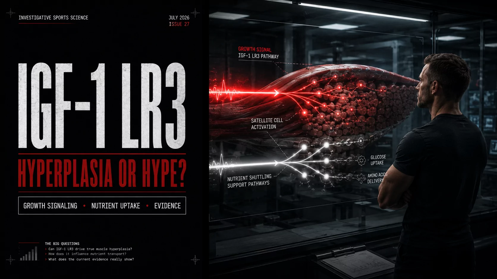
Muscle growth is often described as a simple equation: train hard, consume enough protein, recover, and gradually build larger muscle fibers. At the cellular level, however, the process is considerably more complex. Growth factors, mechanical tension, amino acids, energy availability, satellite cells, and multiple signaling pathways all influence how skeletal muscle responds to training.
Insulin-like growth factor-1, commonly abbreviated as IGF-1, occupies an important place within this network. It helps regulate cellular growth, survival, protein turnover, tissue repair, and metabolic activity. IGF-1 LR3 is a laboratory-engineered analogue designed to preserve IGF-1 receptor activity while interacting less strongly with the binding proteins that normally regulate natural IGF-1.
Those properties have made IGF-1 LR3 a recurring subject in bodybuilding discussions, especially around two ambitious claims: muscle hyperplasia, or an increase in the number of muscle fibers, and nutrient shuttling, the movement and utilization of glucose and amino acids within metabolically active tissues.
The underlying biology is real, but the popular interpretation frequently runs ahead of the evidence. IGF-1 signaling can promote muscle protein synthesis, influence glucose uptake, support satellite-cell activity, and participate in tissue remodeling. That does not prove that externally administered IGF-1 LR3 reliably creates new muscle fibers or directs every additional calorie into muscle in healthy adult athletes.
Educational notice: IGF-1 LR3 has not been approved as a bodybuilding or athletic-performance treatment. This article reviews mechanisms, research findings, limitations, risks, and anti-doping status. It does not provide instructions for administering the compound.
What Is IGF-1 LR3?
IGF-1 LR3, also written as Long R3 IGF-1 or LR3-IGF-1, is a modified analogue of human IGF-1. To understand why the analogue attracts attention, it is useful to begin with the natural hormone.
Natural IGF-1 and the growth hormone axis
Growth hormone is secreted by the pituitary gland in pulses. One of its major systemic effects is stimulating the liver to produce IGF-1. Other tissues, including skeletal muscle, can also produce IGF-1 locally, allowing it to act near the cells that release it.
This distinction matters. Circulating IGF-1 functions as an endocrine signal traveling through the bloodstream, while locally produced IGF-1 can act through autocrine and paracrine signaling within a tissue. Muscle adaptation therefore cannot be reduced to a single blood test or to liver-derived IGF-1 alone.
Once IGF-1 reaches a responsive cell, it can bind to the IGF-1 receptor, or IGF-1R. This receptor is found in numerous tissues rather than exclusively in skeletal muscle. Its wide distribution helps explain both the scientific interest in IGF-1 and the risks of exposing the entire body to a strong growth signal.
How LR3 differs from native IGF-1
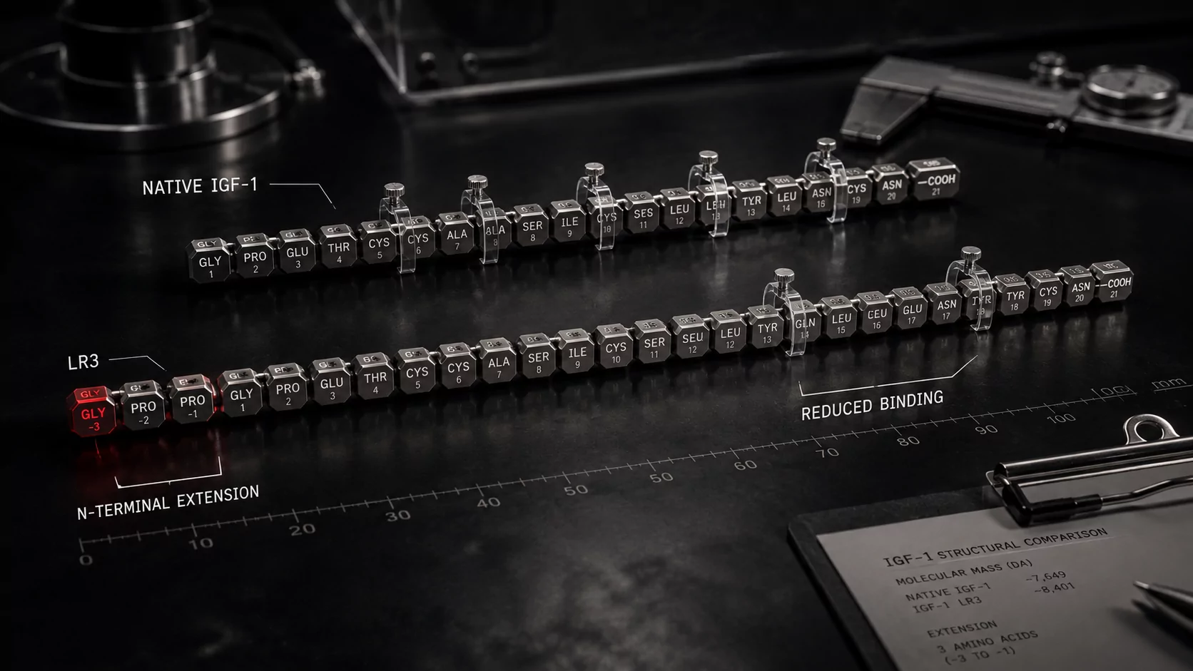
Native human IGF-1 contains 70 amino acids. IGF-1 LR3 contains a 13-amino-acid extension at its N-terminus and replaces the glutamic acid at position three with arginine. These alterations reduce the analogue’s affinity for several IGF-binding proteins.
IGF-binding proteins are not inactive packaging material. They regulate IGF transport, distribution, availability, receptor access, and degradation. An analogue that binds to them less effectively may remain more biologically available in experimental systems, although its behavior can vary with species, tissue, route of exposure, and study design.
Readers researching commercial descriptions of Insulin-like Growth Factor-1 LR3 should distinguish product-page claims from clinical evidence. A listing can describe a compound’s identity or presentation, but it cannot establish that the product is approved, safe, authentic, or proven to deliver a particular bodybuilding result.
Key distinction: IGF-1 LR3 is not simply “more natural IGF-1.” It is a modified research analogue with altered binding-protein interactions and no approved bodybuilding indication.
How IGF-1 Signaling Works Inside Muscle
The appeal of IGF-1 LR3 begins with the signaling network activated after IGF-1R stimulation. This network influences protein synthesis, cellular survival, glucose metabolism, gene expression, and cell proliferation. Two branches are especially relevant: PI3K-Akt signaling and the Ras-Raf-MEK-ERK pathway, often grouped under the broader MAPK system.
IGF-1R: the cellular starting point
IGF-1R is a receptor tyrosine kinase. When an appropriate ligand binds, the receptor changes shape and phosphorylates specific intracellular proteins. These proteins relay the message into the cell rather than allowing the hormone itself to enter and directly “build” tissue.
The downstream response is context-dependent. A mature muscle fiber, a satellite cell, a fibroblast, and a cell in another organ may not respond identically, even when the same receptor is involved. Training status, energy availability, inflammatory signals, local growth factors, insulin sensitivity, and other variables can all modify the outcome.
The PI3K-Akt-mTOR pathway
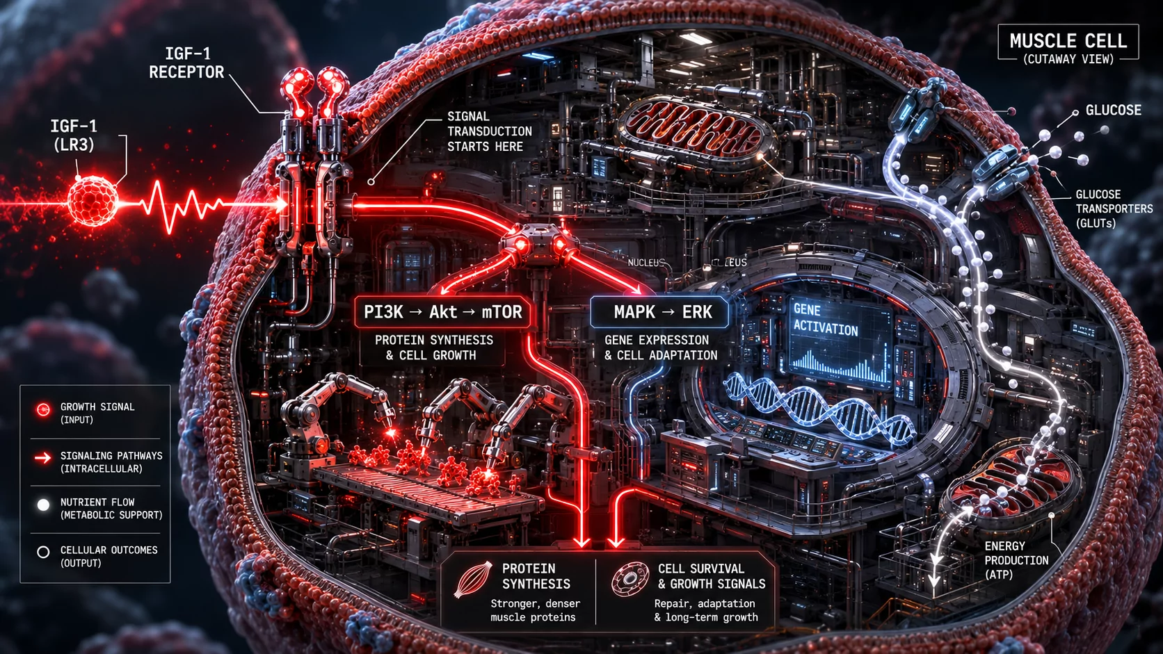
One of the best-known branches begins with phosphoinositide 3-kinase, or PI3K, followed by Akt and downstream regulators that influence mechanistic target of rapamycin complex 1, commonly called mTORC1.
For athletes, the simplified sequence is:
- IGF-1R receives a growth-related signal.
- PI3K and Akt transmit part of that signal inside the cell.
- Downstream regulators change the balance between anabolic and catabolic processes.
- mTORC1 helps coordinate translation and muscle protein synthesis when amino acids and energy are available.
This does not mean IGF-1 is an on-off switch for mTOR. Mechanical loading and amino acids can regulate mTOR-related processes through pathways that are not entirely dependent on IGF-1. Resistance training therefore remains the primary stimulus that tells muscle where and why adaptation is required.
For a broader look at cellular energy production and training recovery, see NAD+ for Athletes: How This Coenzyme Supercharges Mitochondrial Function.
Akt and protein breakdown
Akt signaling may also affect proteins involved in proteolysis. For example, it can influence FOXO transcription factors, which participate in the expression of components involved in the ubiquitin-proteasome system and autophagy.
The net result of IGF-1 signaling may therefore involve both sides of protein balance: encouraging synthesis while modifying pathways associated with breakdown. Nevertheless, whole-body muscle gain still depends on training, nutrition, recovery, endocrine health, and time. A cellular signal does not override those requirements.
MAPK signaling and cell proliferation
The MAPK branch, including ERK signaling, is more closely associated with changes in gene expression, proliferation, differentiation, and cellular responses to environmental stimuli. This pathway is particularly relevant when discussing satellite cells and the hyperplasia hypothesis.
Importantly, proliferation is not synonymous with muscle-fiber creation. A precursor cell can divide without ultimately becoming a functional new muscle fiber. It may remain in the stem-cell pool, differentiate, fuse into an existing fiber, undergo programmed cell death, or respond differently depending on the surrounding tissue environment.
Hypertrophy Versus Hyperplasia
Few bodybuilding terms generate as much confusion as hyperplasia. It is frequently used as a more impressive synonym for muscle growth, even though it describes a different biological event from hypertrophy.
What is muscle hypertrophy?
Hypertrophy is an increase in the size of existing muscle fibers. A muscle fiber is a large, multinucleated cell. With progressive resistance training and adequate recovery, its contractile machinery and supporting cellular structures can expand, increasing the fiber’s cross-sectional area.
This is the dominant, well-supported model used to explain measurable muscle growth in adult humans. Hypertrophy can involve:
- Greater synthesis and accumulation of contractile proteins
- Expansion of cellular components that support force production
- Changes in glycogen, water, enzymes, and other non-contractile elements
- Addition of myonuclei through satellite-cell fusion under certain conditions
- Remodeling of extracellular matrix and connective tissue
What is muscle hyperplasia?
Hyperplasia means an increase in the number of cells or, in this context, muscle fibers. The concept is established in some tissues and has been observed under specific experimental conditions in certain animal models. Whether substantial muscle-fiber hyperplasia commonly contributes to adult human resistance-training adaptation remains unresolved.
Researchers face a difficult measurement problem. A larger muscle does not automatically contain more fibers. To prove whole-muscle hyperplasia, investigators would need to count fibers reliably across the relevant muscle before and after an intervention without confusing fiber splitting, changes in muscle architecture, sampling error, or differences in cross-sectional area.
Does IGF-1 LR3 promote hyperplasia?
IGF-1-related signaling creates a biologically plausible route through which precursor-cell proliferation and muscle regeneration could be enhanced. Experimental work shows that IGF-1 can activate satellite cells and influence their proliferation and differentiation. Animal and cell-culture research also demonstrates that IGF-1 can increase muscle size or affect regenerative activity.

What has not been established is that IGF-1 LR3 reliably produces a meaningful increase in whole-muscle fiber number in healthy adult bodybuilders. Most direct LR3 experiments have involved cells, livestock, rodents, or other animal models. Findings from those systems can reveal mechanisms, but they cannot be translated into guaranteed human outcomes.
Evidence-based conclusion: The claim that IGF-1 LR3 “causes hyperplasia” should be treated as a hypothesis derived from growth-factor biology and preclinical research—not as a demonstrated bodybuilding outcome in humans.
Fiber splitting is not automatically hyperplasia
Some discussions describe enlarged fibers splitting into smaller daughter fibers. Histological observations that resemble splitting have been reported in selected animal models and extreme loading conditions. However, identifying split-looking fibers in a tissue section does not necessarily prove the creation of fully independent, functional fibers across an entire muscle.
Orientation, branching, regeneration, injury, and sampling methods can affect what appears in a microscopic image. This is one reason responsible interpretation requires more than a dramatic before-and-after illustration.
Satellite Cells: The More Defensible Part of the Muscle-Growth Story
Satellite cells are muscle-resident stem cells positioned between a muscle fiber’s plasma membrane and its surrounding basal lamina. They are normally relatively inactive, but they can respond to mechanical loading, injury, inflammatory signals, and local growth factors.
Activation, proliferation, and differentiation
When activated, satellite cells can pass through several stages:
- Activation: The cell leaves its resting state in response to local signals.
- Proliferation: It divides to create a larger population of precursor cells.
- Differentiation: Some descendants begin developing a muscle-specific identity.
- Fusion: Differentiated cells may fuse with an existing fiber or participate in the formation of new myotubes during regeneration.
- Self-renewal: A portion returns to a stem-like state, preserving the satellite-cell pool.
IGF-1 can influence several parts of this process. A review of IGF-1 signaling in skeletal muscle describes roles in protein metabolism, regeneration, and satellite-cell activation. Interested readers can review the underlying mechanisms in this peer-reviewed overview indexed by the National Library of Medicine.
Myonuclear addition and hypertrophy
Satellite-cell fusion can add nuclei to an existing muscle fiber. These nuclei are known as myonuclei. Because each nucleus supports gene expression across a finite region of the fiber, adding myonuclei may help an enlarging fiber manage the demands of maintaining more cellular volume.
This gives IGF-1-related satellite-cell activity a credible role in hypertrophy without requiring the creation of entirely new fibers. A muscle can become significantly larger because its existing fibers grow and gain nuclear support.
Regeneration is not the same as routine growth
Satellite cells are indispensable for repairing severe muscle damage, but normal resistance-training adaptation does not require athletes to repeatedly destroy muscle tissue. Productive training applies enough tension and fatigue to stimulate adaptation while managing damage so performance can recover.
Growth-factor signaling that assists regeneration should therefore not be interpreted as permission to pursue excessive soreness or injury. For a separate discussion of peptide-related repair claims, see BPC 157 Explained: How This Peptide Accelerates Injury Recovery.
What “Nutrient Shuttling” Actually Means
Nutrient shuttling is a bodybuilding term rather than a single, precisely defined physiological process. In practice, it usually refers to directing glucose, amino acids, and other nutrients toward skeletal muscle for energy production, glycogen restoration, repair, and growth.
The concept overlaps with the scientific term nutrient partitioning: how the body distributes and uses incoming energy among muscle, liver, adipose tissue, and other organs.
IGF-1 has insulin-like metabolic effects
IGF-1 received its name because it shares structural and functional similarities with insulin. The IGF-1 receptor and insulin receptor are related, and their signaling networks overlap. IGF-1 can influence glucose uptake and metabolism, although insulin remains the principal acute regulator of blood glucose after meals.
After receptor activation, PI3K-Akt-related signaling may promote the movement of glucose transport machinery toward the cell membrane in responsive tissues. This can facilitate glucose entry into cells, where it may be:
- Used to produce ATP
- Stored as glycogen
- Processed through glycolysis
- Directed into other metabolic pathways
This helps explain both the attraction and the danger. A signal that increases glucose disposal may contribute to lower circulating glucose. Hypoglycemia is not evidence that a compound is “working perfectly”; it is a potentially serious adverse effect.
Glucose uptake is not muscle-selective
A common marketing image depicts nutrients bypassing fat cells and flowing exclusively into trained muscles. Human metabolism is not that selective. Systemic IGF-1 receptor activation can affect multiple tissues, and nutrient distribution remains influenced by:
- Energy balance
- Insulin sensitivity
- Muscle contractions and recent training
- Glycogen depletion
- Blood flow
- Meal composition
- Adipose-tissue biology
- Genetics and hormonal status
Resistance exercise itself improves glucose uptake by working muscle through contraction-mediated mechanisms. That is one reason training before an appropriate recovery meal can produce effective nutrient partitioning without relying on an experimental growth-factor analogue.
Glycogen restoration and the “full” appearance
Muscle glycogen is stored with water. When depleted muscle restores glycogen, the tissue can look and feel fuller. This visual change is meaningful for training readiness and physique presentation, but it is not identical to the addition of new contractile protein.
Bodybuilders should separate at least three outcomes:
- Glycogen and water restoration: Relatively rapid and responsive to carbohydrate intake
- Muscle protein remodeling: Slower and dependent on repeated training-recovery cycles
- New muscle-fiber formation: Unproven as a meaningful LR3-driven outcome in adult humans
Amino acids and protein synthesis
Amino acids provide the raw materials for protein synthesis, while essential amino acids—especially leucine—also function as nutritional signals. IGF-1-related pathways may create a cellular environment favorable to anabolism, but amino acids must still be present.
More signaling does not manufacture protein from nothing. If energy intake, dietary protein, micronutrient status, sleep, or training quality is inadequate, no growth factor can fully compensate.
For another perspective on substrate use and metabolic claims in bodybuilding, read L-Carnitine Explained: More Than Just a Fat Burner for Bodybuilders.
Nutrient shuttling versus fat loss
Improved glucose uptake should not be confused with guaranteed fat loss. Fat gain or loss is strongly governed by long-term energy balance, although nutrient partitioning affects where and how energy is stored and used.
An athlete consuming a sustained calorie surplus can still gain fat. Similarly, a compound that changes short-term glucose dynamics does not suspend the laws of energy balance. Training status and insulin sensitivity may influence the quality of weight gain, but neither creates unlimited muscle-building capacity.
Recovery, Protein Turnover, and Tissue Remodeling
Recovery is often framed as the disappearance of soreness. Biologically, it is a coordinated period during which muscle proteins turn over, glycogen is replenished, damaged cellular structures are managed, inflammation is regulated, and the nervous system regains performance capacity.
Muscle protein synthesis
IGF-1 signaling can support muscle protein synthesis through Akt-mTOR-related mechanisms. In experimental models, it can also oppose atrophy-related pathways. Yet the magnitude of adaptation is determined by the complete environment around that signal.
Mechanical tension tells the tissue that greater force-producing capacity is needed. Dietary protein supplies amino acids. Adequate energy supports expensive biosynthetic processes. Sleep and recovery allow repeated high-quality training. IGF-1 is one participant within this system, not an independent replacement for it.
Connective tissue and extracellular matrix
Muscle fibers operate within an extracellular matrix that transfers force and supports tissue architecture. Growth-factor signaling can affect fibroblasts, collagen turnover, blood vessels, and other components involved in remodeling.
This broader activity is not automatically beneficial. Excessive or poorly regulated growth signaling may produce maladaptive changes as well as desired ones. Tissue growth must be coordinated; making one element grow faster does not guarantee that tendons, nerves, blood vessels, and joint structures will adapt in perfect proportion.
Recovery signals can have systemic consequences
Because IGF-1 receptors are distributed throughout the body, the idea of a purely muscle-specific systemic effect is unrealistic. Experimental research has reported effects on organs and non-muscle tissues in animal models. That does not predict an exact outcome in humans, but it reinforces the need to avoid interpreting the compound as a targeted recovery supplement.
IGF-1 LR3 Versus Growth Hormone
Growth hormone and IGF-1 belong to the same physiological axis, but they are not interchangeable. Growth hormone acts through its own receptor and produces both direct effects and indirect effects mediated partly through IGF-1.
How growth hormone acts
Growth hormone influences liver-derived IGF-1, fat metabolism, fluid balance, connective-tissue turnover, and numerous other processes. Its release is naturally pulsatile, with sleep, exercise, nutritional state, age, and other factors affecting secretion.
Growth hormone secretagogues, such as those discussed in CJC-1295 No DAC and Ipamorelin, aim to stimulate parts of the body’s own GH-release machinery. That mechanism differs from directly exposing IGF-1 receptors to an external analogue.
How IGF-1 LR3 differs
IGF-1 LR3 acts primarily as an IGF-1 receptor agonist with reduced affinity for IGF-binding proteins. It does not reproduce the full pattern of growth hormone action, nor does it preserve the same physiological feedback and tissue-distribution characteristics as naturally generated IGF-1.
| Feature | Growth hormone | IGF-1 LR3 |
|---|---|---|
| Primary receptor | Growth hormone receptor | IGF-1 receptor |
| Relationship to IGF-1 | Can stimulate IGF-1 production | Modified IGF-1 analogue |
| Natural secretion pattern | Pulsatile | No natural LR3 secretion |
| Binding-protein interaction | Indirectly affects the IGF system | Engineered for reduced IGFBP affinity |
| Approved bodybuilding use | None | None |
| Competitive sport | Prohibited under anti-doping rules | IGF-1 analogues are prohibited |
Readers comparing the two should also distinguish LR3 from pharmaceutical recombinant human growth hormone products discussed in The Truth About Dragontropin: Is This HGH Brand Worth Your Money?. Product identity, pharmacology, evidence, and safety cannot be transferred from one hormone or analogue to another.
What the Scientific Evidence Actually Supports
The strongest conclusions come from separating evidence into levels rather than merging every study into a single promise.
Established IGF-1 biology
There is strong evidence that natural IGF-1:
- Binds to IGF-1R and activates intracellular signaling
- Influences Akt, mTOR, MAPK, and related networks
- Participates in skeletal-muscle growth and regeneration
- Affects satellite-cell behavior
- Influences protein turnover and glucose metabolism
- Acts in numerous tissues outside skeletal muscle
What preclinical LR3 research shows
IGF-1 LR3 has been used extensively as a laboratory tool because reduced binding-protein affinity can make IGF-1 receptor effects easier to study. Experiments in cells and animals have investigated glucose uptake, protein metabolism, fetal development, organ growth, neurobiology, and tissue-specific signaling.
These studies confirm that LR3 is biologically active. They do not establish a safe or effective performance-enhancement protocol for healthy adults.
What human exercise research suggests
Human research supports a relationship between exercise, local IGF-1 biology, and satellite-cell responses. For example, investigators have observed increased association of IGF-1 with satellite cells after resistance exercise. This helps demonstrate that IGF-1 participates in normal adaptation.
It does not demonstrate that injecting a modified analogue produces the same spatial, temporal, or physiological signal. Locally generated IGF-1 after training may act in a controlled microenvironment that differs substantially from systemic exposure.
What remains unproven
High-quality human evidence does not currently establish that IGF-1 LR3:
- Reliably increases total muscle-fiber number in trained adults
- Selectively shuttles nutrients only into skeletal muscle
- Produces permanent muscle gains independent of training and diet
- Is safe for non-medical physique enhancement
- Has a validated bodybuilding dose, cycle length, or stacking protocol
- Can be used without clinically meaningful hypoglycemic or proliferative risk
IGF-1 LR3 Evidence Ladder
- Strongest: Basic IGF-1 receptor and signaling biology
- Moderate: Natural IGF-1 involvement in muscle adaptation and regeneration
- Preclinical: LR3 effects in cell culture and animal models
- Speculative: Reliable whole-muscle hyperplasia in adult bodybuilders
- Unsupported: Guaranteed lean gains with no systemic risk
Potential Risks and Side Effects
The same properties that make IGF-1 biologically interesting also create risk. It influences glucose metabolism, cellular survival, tissue growth, and proliferation. These are not minor systems to manipulate without medical oversight.
Hypoglycemia
Low blood glucose is one of the most immediate concerns associated with pharmacological IGF-1 activity. Possible symptoms include:
- Shaking or sweating
- Sudden hunger
- Dizziness or weakness
- Headache
- Rapid heartbeat
- Blurred vision
- Confusion or abnormal behavior
- Loss of consciousness or seizure in severe cases
Severe hypoglycemia is a medical emergency. The risk can become more difficult to predict when an unapproved product has uncertain identity, purity, concentration, or storage history.
Fluid retention and soft-tissue symptoms
IGF-related therapies may be associated with swelling, soft-tissue changes, headaches, or musculoskeletal discomfort. Rapid changes in tissue volume can also create numbness or nerve-compression symptoms in susceptible individuals.
Abnormal tissue and organ growth
IGF-1 receptors exist in organs, connective tissue, and other cell populations. A systemic growth signal cannot be assumed to enlarge skeletal muscle alone. Preclinical studies showing effects on non-muscle tissues are a warning against the phrase “targeted muscle growth.”
Cell-proliferation concerns
IGF-1 signaling can promote cellular survival and proliferation. That does not mean IGF-1 LR3 automatically causes cancer, and simplistic claims in either direction should be avoided. The legitimate concern is that enhancing a growth-and-survival pathway may be inappropriate in someone with an existing or undiagnosed neoplastic process.
The current FDA prescribing information for pharmaceutical mecasermin—an approved recombinant human IGF-1 medicine, not LR3—contains substantial warnings and monitoring requirements. It can be reviewed in the official FDA label for Increlex. The existence of an approved IGF-1 medication does not validate non-medical use of LR3; instead, the label illustrates how seriously medical IGF-1 exposure must be managed.
Unknown product quality
Products sold outside regulated pharmaceutical distribution may be mislabeled, degraded, contaminated, under-strength, over-strength, or substituted with another substance. A professional-looking vial or third-party report does not eliminate the full chain-of-custody problem.
Peptide and protein products are also sensitive to manufacturing and storage conditions. Purity by one analytical method does not automatically confirm sterility, correct folding, biological activity, endotoxin control, or accurate concentration.
Seek urgent medical care for loss of consciousness, seizure, severe confusion, chest pain, major breathing difficulty, rapidly worsening swelling, a serious allergic reaction, or signs of severe infection. Anyone experiencing suspected hypoglycemia should treat it as a medical issue rather than a routine training side effect.
Medical Approval and Competitive-Sport Status
IGF-1 LR3 is not approved for bodybuilding
The United States has an approved recombinant human IGF-1 medication called mecasermin for narrowly defined pediatric growth disorders. IGF-1 LR3 is a different molecule and does not have an FDA-approved indication for muscle gain, recovery, anti-aging, or athletic performance.
“Research use” labeling should not be mistaken for medical authorization. It generally indicates that a material is marketed for laboratory work rather than approved treatment in humans.
IGF-1 analogues are prohibited in tested sport
IGF-1 and its analogues are prohibited under World Anti-Doping Agency rules. The prohibition applies in and out of competition under the relevant category. Tested athletes should consult the current list directly because anti-doping rules are updated periodically.
The 2026 WADA Prohibited List includes IGF-1 among prohibited growth factors and related substances. Athletes remain responsible for substances detected in their samples, including those arising from contaminated or incorrectly labeled products.
Common IGF-1 LR3 Myths
Myth 1: “IGF-1 LR3 creates brand-new muscle fibers”
Reality: IGF-1 signaling can influence satellite cells, proliferation, differentiation, and regeneration. Direct evidence of meaningful LR3-induced whole-muscle hyperplasia in healthy adult humans is lacking.
Myth 2: “Every nutrient goes directly into muscle”
Reality: IGF-1-related signaling can affect glucose metabolism, but systemic exposure is not restricted to skeletal muscle. Energy balance, training, insulin sensitivity, glycogen status, and tissue-specific biology continue to govern nutrient partitioning.
Myth 3: “It works like insulin without insulin’s risks”
Reality: IGF-1 and insulin are distinct hormones, but their signaling networks overlap. IGF-1 activity can still lower blood glucose and create clinically significant hypoglycemic risk.
Myth 4: “More receptor activation means more muscle”
Reality: Biological systems have feedback loops, saturation points, competing pathways, and adverse effects. Increasing a systemic signal does not produce unlimited, muscle-specific growth.
Myth 5: “Longer activity automatically makes LR3 superior”
Reality: Prolonged biological exposure may increase convenience in a laboratory experiment, but it can also extend adverse metabolic and proliferative effects. Longer is not synonymous with safer or better.
Myth 6: “A laboratory purity result proves the product is safe”
Reality: Purity testing is only one element of pharmaceutical quality. It does not necessarily establish sterility, potency, identity across every vial, correct storage, absence of endotoxin, or suitability for human use.
What Athletes Can Learn from the Biology Without Using LR3
The science of IGF-1 reinforces several practical principles that do not require an experimental compound.
Progressive loading creates the tissue-specific signal
Muscle adapts where mechanical demand is repeatedly imposed. A well-designed program supplies enough volume and intensity to stimulate adaptation while allowing performance to recover.
Carbohydrates support training and glycogen restoration
Appropriate carbohydrate intake can restore glycogen and support high-quality training. The amount required depends on training volume, body size, goals, total calories, and individual tolerance.
Protein distribution supports repeated synthesis opportunities
Adequate daily protein matters more than chasing a narrow anabolic window. Distributing high-quality protein across several meals can provide repeated essential-amino-acid exposure during the day.
Sleep supports endocrine and metabolic health
Consistent sleep assists recovery, glucose regulation, appetite control, training performance, and the natural GH-IGF axis. Poor sleep can undermine nutrient partitioning even when the diet looks optimal on paper.
Insulin sensitivity is trainable
Resistance exercise, aerobic activity, maintaining a suitable body-fat range, adequate sleep, and a minimally processed diet can improve metabolic health. These fundamentals influence nutrient use more reliably than speculative claims about selective shuttling.
Frequently Asked Questions
No. It is a modified analogue with a 13-amino-acid N-terminal extension and an arginine substitution at position three. These changes reduce its affinity for IGF-binding proteins and alter its behavior relative to native IGF-1.
No. The mechanism is biologically plausible at the level of cell proliferation and regeneration, but substantial muscle-fiber hyperplasia has not been demonstrated as a reliable outcome in healthy adult humans using LR3.
IGF-1 receptor signaling is anabolic in multiple experimental contexts and can support protein synthesis and satellite-cell activity. However, there is insufficient controlled human evidence to define the muscle-building effectiveness or safety of IGF-1 LR3 for bodybuilding.
It refers broadly to the uptake, storage, and use of nutrients such as glucose and amino acids. The phrase is often used more confidently than the science allows. Nutrient distribution is controlled by many factors and is not made exclusively muscle-specific by IGF-1 signaling.
There is no sound basis for calling it a safe alternative. The compounds differ, but IGF-1 activity can still disrupt glucose control and cause hypoglycemia. LR3 also raises broader concerns related to systemic growth signaling and unregulated product quality.
IGF-1 LR3 is not FDA-approved for bodybuilding, athletic performance, recovery, or anti-aging. Pharmaceutical mecasermin is a different form of recombinant human IGF-1 approved for specific pediatric growth disorders under medical supervision.
Yes. IGF-1 and its analogues are prohibited under WADA rules. Competitive athletes should verify the current Prohibited List and obtain qualified anti-doping guidance before using any medication or supplement.
Growth hormone activates the growth hormone receptor and can stimulate IGF-1 production while producing other direct metabolic effects. IGF-1 LR3 directly activates IGF-1-related signaling as a modified analogue. One is not a simple substitute for the other.
Final Verdict: Promising Mechanisms, Unproven Bodybuilding Claims
IGF-1 is undeniably important to muscle biology. It helps regulate protein turnover, supports satellite-cell activity, participates in regeneration, and influences glucose metabolism. IGF-1 LR3 was engineered to reduce binding-protein interactions, making it a potent experimental tool for investigating those processes.
That science provides a plausible explanation for why LR3 could affect muscle growth and nutrient handling. It does not prove the most aggressive bodybuilding claims attached to it.
Hypertrophy of existing fibers remains the best-supported explanation for adult human muscle growth. Satellite-cell activation and myonuclear addition can contribute to that process without requiring true hyperplasia. Meanwhile, the phrase “nutrient shuttling” should be understood as a network of metabolic effects—not as a guarantee that calories will bypass fat storage and flow exclusively into muscle.
The most accurate conclusion is therefore balanced: IGF-1 LR3 activates pathways relevant to anabolism, regeneration, and glucose utilization, but reliable muscle-fiber hyperplasia, selective nutrient partitioning, and safe performance enhancement have not been demonstrated in healthy adult humans.
For athletes, the gap between an interesting mechanism and a proven outcome matters. It is the difference between understanding the science and being persuaded by a promise.